Step-by-Step Study Material
STEP 1: Understanding the Problem
The Core Challenge
1% of all live births → congenital heart defect
Every defect is UNIQUE
Every child's anatomy is DIFFERENT
Why This Is Difficult
| Traditional Approach | The Problem |
|---|---|
| Off-the-shelf devices | Not designed for specific children |
| Manual 3D modeling | Takes ~4 hours per case |
| Standard imaging only | Limited anatomical understanding |
| Trial during surgery | Risk of failed procedures |
Real-World Consequence
A child underwent two failed heart repair surgeries because surgeons could not locate the defect using traditional methods
Key Insight: Surgeons needed to understand the exact anatomy before entering the operating room
STEP 2: The Solution Framework — Three Core Needs
Before learning the tools, understand what problems needed solving:
NEED 1: See the heart accurately
         ↓
NEED 2: Model it quickly enough for clinical use
         ↓
NEED 3: Simulate what happens BEFORE surgery
STEP 3: The Technology Stack — Layer by Layer
Layer 1 — Medical Imaging Input
Sources of data already available to care teams:
- CT scans
- MRI
- 3D Ultrasound (echocardiography)
These images existed before AI — the challenge was processing them fast enough and precisely enough
Layer 2 — 3D Slicer & SlicerHeart
What it is:
- 3D Slicer = open source software for visualizing and analyzing 3D medical images
- SlicerHeart = an extension built specifically for pediatric hearts
What it does:
Raw medical images → Segmented, visualized 3D heart model
Who built it: Dr. Matthew Jolley's lab at CHOP, with open source community contributions from Stanford and Boston Children's Hospital
Layer 3 — MONAI + Auto3DSeg (The AI Acceleration Layer)
What is MONAI?
- Open source medical imaging AI framework
- Co-founded by NVIDIA
What is Auto3DSeg?
- NVIDIA's automated 3D segmentation implementation within MONAI
- Trained on pairs of prior images and manually created models
The Machine Learning Workflow:
Step 1: Collect 10–20 image-model pairs (human-made)
Step 2: Train a segmentation network on those pairs
Step 3: Apply the trained model to new patient images
Step 4: Output meets same quality as human expert
The Impact:
| Before AI | After AI |
|---|---|
| 4 hours per model | Seconds |
| Skilled researcher required | Automated |
| Research use only | Routine clinical use |
"Machine learning has become just bread and butter" — Dr. Jolley
Layer 4 — Newton & NVIDIA Warp (The Simulation Layer)
What is NVIDIA Warp?
- A Python framework that runs physics simulations on GPUs
What is Newton?
- An open source physics engine built on Warp
- Originally designed for robot training simulations
- Now being adapted for biomechanical cardiac simulation
What does this layer solve?
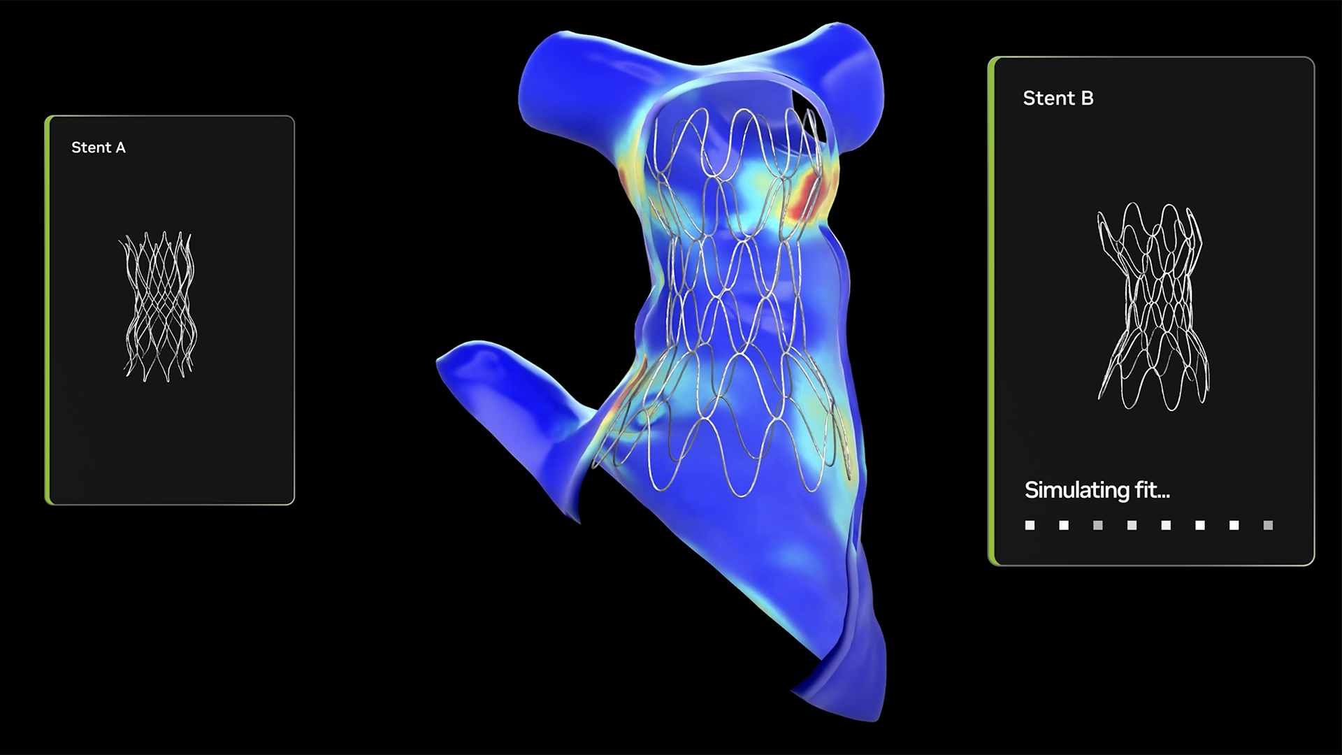
Visualization answers: "What does the heart look like?"
Simulation answers: "What will HAPPEN when we put a device inside it?"
3D Heart Model + Device Specifications
            ↓
     Newton/Warp Simulation
            ↓
   Predicted device behavior in THIS child's anatomy
Time improvement:
| Before GPU Simulation | After GPU Simulation |
|---|---|
| 4 hours or overnight | Near real-time |
| One configuration tested | Multiple configurations compared |
Clinical Application:
- Closure devices (sealing holes in hearts)
- Transcatheter valve simulations (future goal)
- Same-day decision making becomes possible
Layer 5 — OpenUSD & NVIDIA Omniverse (The Interaction Layer)
What is OpenUSD?
- Open source 3D interoperability standard
- Allows diverse data and solvers to be combined
What is NVIDIA Omniverse?
- Digital twin platform
What this enables:
Patient Images → 3D Simulation → Virtual Reality Environment
                                          ↓
                          Clinician interacts using natural language
                          (Vision-Language Models / VLMs)
                                          ↓
                          "Show me how Device A fits vs Device B"
This layer is currently in development — it represents the future direction
STEP 4: The Complete Workflow — End to End
PATIENT ARRIVES
      ↓
Existing images collected (CT, MRI, Ultrasound)
      ↓
MONAI + Auto3DSeg segments the anatomy (seconds)
      ↓
SlicerHeart generates precise 3D heart model
      ↓
Newton/Warp simulates device deployment
      ↓
Clinician compares device options in near real-time
      ↓ (future)
VR environment with language-model interaction
      ↓
INFORMED SURGICAL DECISION — before anyone enters the OR
STEP 5: Why Open Source Is the Critical Enabler
The Economics Problem
~2.4 million people in U.S. have congenital heart disease
Each case is unique → no standardized solution
Small market → traditional companies won't invest
No single institution can build everything alone
How Open Source Solves This
| Traditional Commercial Model | Open Source Model |
|---|---|
| Company funds development | Community shares development |
| Profit motive required | Research + philanthropy driven |
| Tools locked behind licenses | Free to use and build upon |
| One institution's resources | Many institutions' combined expertise |
The Collaboration Network
- CHOP — core development, clinical application
- Boston Children's Hospital — 500+ modeled surgeries/year, tool contributions
- Stanford — additional tool development
- National consortium — forming to build next-generation shared infrastructure
- NVIDIA — maintains industrial-scale open platforms
"Open source defies traditional economics for small and heterogeneous populations" — Dr. Jolley
STEP 6: Measuring Real-World Impact
Current Scale
| Institution | Impact |
|---|---|
| CHOP | ~200 modeled cases/year (growing) |
| Boston Children's | 500+ cases/year, >50% of all cardiac surgeries |
| U.S. hospitals using cardiac modeling | 20+ |
Clinical Milestones
- Ventricular septal defect (hole between heart chambers) modeling is now standard of care at CHOP
- The failed-repair case: 3D modeling led to first-attempt success after two prior failures
- CHOP's IDEA Lab is expanding these tools beyond cardiology to multiple disciplines
Concept Summary Map
CONGENITAL HEART DISEASE (unique per child)
              ↓
         IMAGING DATA
              ↓
    MONAI + Auto3DSeg (AI segmentation)
              ↓
    SlicerHeart / 3D Slicer (3D modeling)
              ↓
    Newton + NVIDIA Warp (physics simulation)
              ↓
    OpenUSD + Omniverse + VLMs (interaction)
              ↓
    SAFER, MORE PRECISE PEDIATRIC CARDIAC CARE
              ↑
    Enabled throughout by OPEN SOURCE collaboration
Key Terms to Remember
| Term | Simple Definition |
|---|---|
| Congenital heart defect | Heart abnormality present from birth |
| Segmentation | AI identifying and outlining specific structures in medical images |
| MONAI | Open source AI framework for medical imaging |
| Auto3DSeg | Automated 3D segmentation tool by NVIDIA |
| SlicerHeart | Open source tool for pediatric heart modeling |
| Newton | Open source physics engine for simulation |
| NVIDIA Warp | GPU-accelerated Python framework for physics |
| OpenUSD | Open standard for 3D data interoperability |
| Digital twin | Virtual replica of a physical object (here: a child's heart) |
| VLM | Vision-Language Model — AI that understands images and natural language |
Self-Check Questions
- Why can't surgeons simply use off-the-shelf devices for children with congenital heart defects?
- What was the workflow before AI, and what made it impractical for routine clinical use?
- What specific role does MONAI play versus Newton in this system?
- Why is open source particularly important for rare pediatric conditions?
- What is the difference between visualization and simulation in this context, and why does that distinction matter clinically?